 |
|  |
| 
Previous issue | Next issue | Archive
![]() Volume 13 (4); December 25, 2023 [Booklet] [EndNote XML for Agris]
Volume 13 (4); December 25, 2023 [Booklet] [EndNote XML for Agris]
 Research Paper
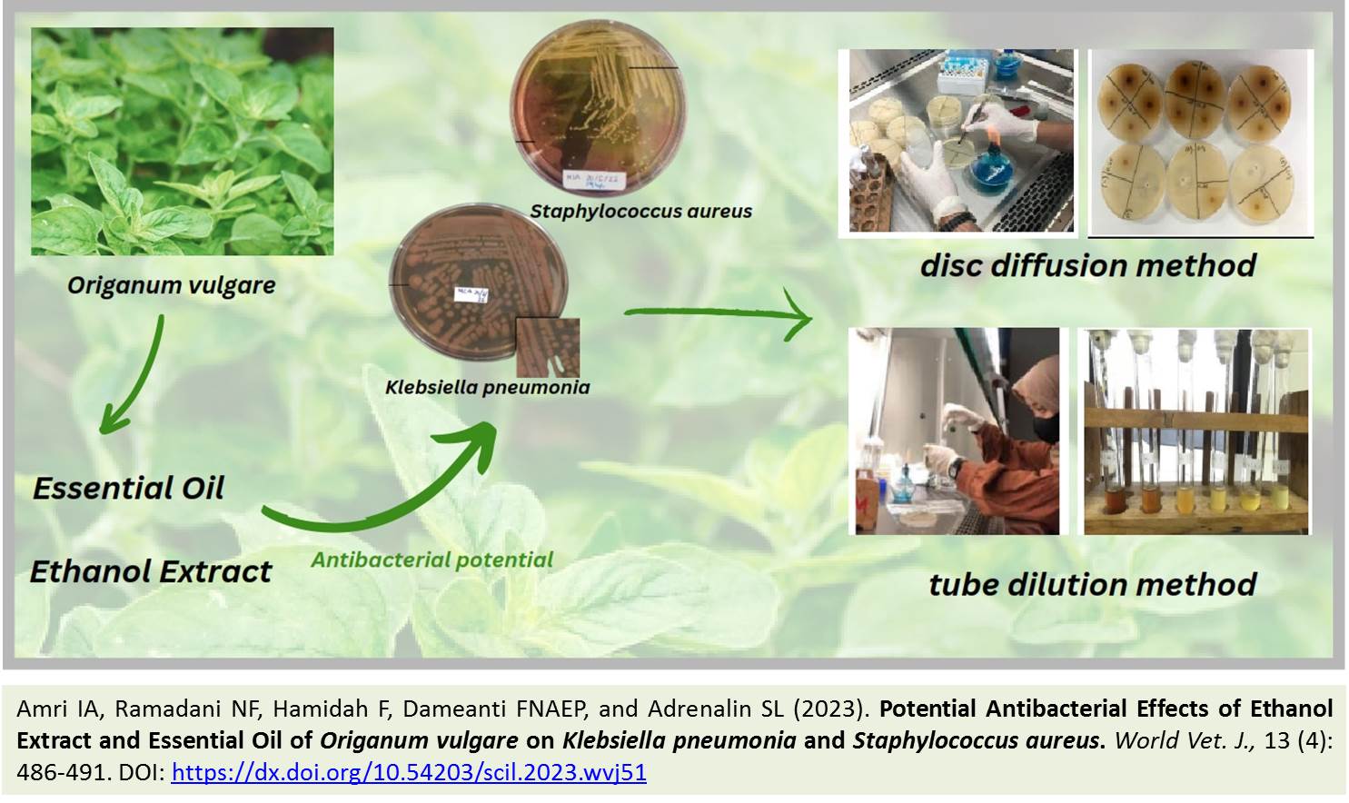
Potential Antibacterial Effects of Ethanol Extract and Essential Oil of Origanum vulgare on Klebsiella pneumonia and Staphylococcus aureus
Amri IA, Ramadani NF, Hamidah F, Dameanti FNAEP, and Adrenalin SL.
World Vet. J. 13(4): 486-491, 2023; pii:S232245682300051-13
DOI: https://dx.doi.org/10.54203/scil.2023.wvj51
ABSTRACT: Klebsiella pneumonia (K. pneumonia) and Staphylococcus aureus (S. aureus) are pathogenic bacteria causing various infectious diseases in humans and animals. Currently, herbal ingredients are widely used as antibacterial agents to combat bacterial infections due to their lower side effects, compared to chemical drugs. One such plant with medicinal promise as an antibacterial agent is the oregano plant (Oregano vulgare). It contains substances, such as tannin, flavonoids, carvacrol, thymol, and saponin. Therefore, the current study was conducted to regularly compare the in vitro antibacterial potential of ethanol extract essential oil oregano (Oregano vulgare) on K. pneumonia and S. aureus. In this research, the diffusion method using discs was employed to observe the inhibition zones, while the dilution tube method was utilized to determine the minimum inhibitory concentration (MIC) of the ethanol extract and essential oil of oregano against the test bacteria. The bacterial treatment group received the test material at concentrations of 100%, 50%, 25%, and 12.5%. The obtained data were analyzed descriptively in terms of zone inhibition and MIC values. According to the disc diffusion test, the essential oil of oregano demonstrated greater efficacy as an antibacterial agent against K. pneumoniae at a concentration of 100%, resulting in an average inhibition zone of 18 mm. Conversely, for S. aureus, a concentration of 1.5% of the essential oil exhibited higher effectiveness, yielding an average inhibition zone of 30 mm. Based on the MIC values, the essential oil was more effective as an antibacterial for K. pneumonia at a concentration of 0.2% (2 mg/mL), while for S. aureus it was more effective at a concentration of 0.19% (1.9 mg/mL).
Keywords: Antibacterial, Ethanol Extract, Essential Oil, Oregano vulgare
[Full text-PDF] [Crossref Metadata] [Scopus] [Export from ePrints]
 Research Paper
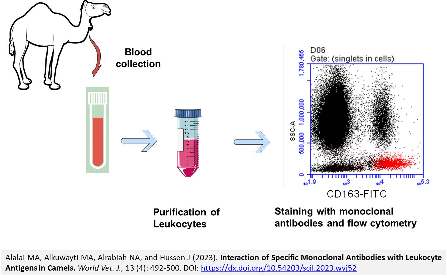
Interaction of Specific Monoclonal Antibodies with Leukocyte Antigens in Camels
Alalai MA, Alkuwayti MA, Alrabiah NA, and Hussen J.
World Vet. J. 13(4): 492-500, 2023; pii:S232245682300052-13
DOI: https://dx.doi.org/10.54203/scil.2023.wvj52
ABSTRACT: The dromedary camel as a livestock species significantly impacts the economy of arid and semi-arid regions worldwide. The identification of cross-reactive antibodies against pivotal immune cell markers acts as a valuable method to investigate the immune system of camels. The aim of the present study was to identify new monoclonal antibodies that react with camel leukocyte subsets using flow cytometry and multicolor immunofluorescence. The expression patterns of the tested antibodies indicated cross-reactivity of the anti-bovine CD9 monoclonal antibody clones LT86A and Hl9a with different binding potential. Although all leukocyte subpopulations stained positively with the CD9 antibodies, monocytes showed the highest CD9 abundance, compared to lymphocytes and granulocytes. No cross-reactivity was identified for the tested monoclonal antibodies against equine CD8a (clone: ETC142BA1), mouse CD3 (clone: CD3-12), human CD3 (clone: T3/2/16A9), human CD206 (clone: MMR), and bovine granulocytes (clone: CH138A). The present study revealed that only camel monocytes showed positive staining with the anti-ovine CD5 mAb (clone ST1), which is in contrast to the human and murine systems. The present findings indicated low homogeneity between camels and other species in the antigenic structure of leukocyte antigens, highlighting the need to develop camel-specific mAbs against the main immune cell markers.
Keywords: Antibodies, Camel, Cell marker, Flow cytometry, Immunity
[Full text-PDF] [Crossref Metadata] [Scopus] [Export from ePrints]
 Research Paper
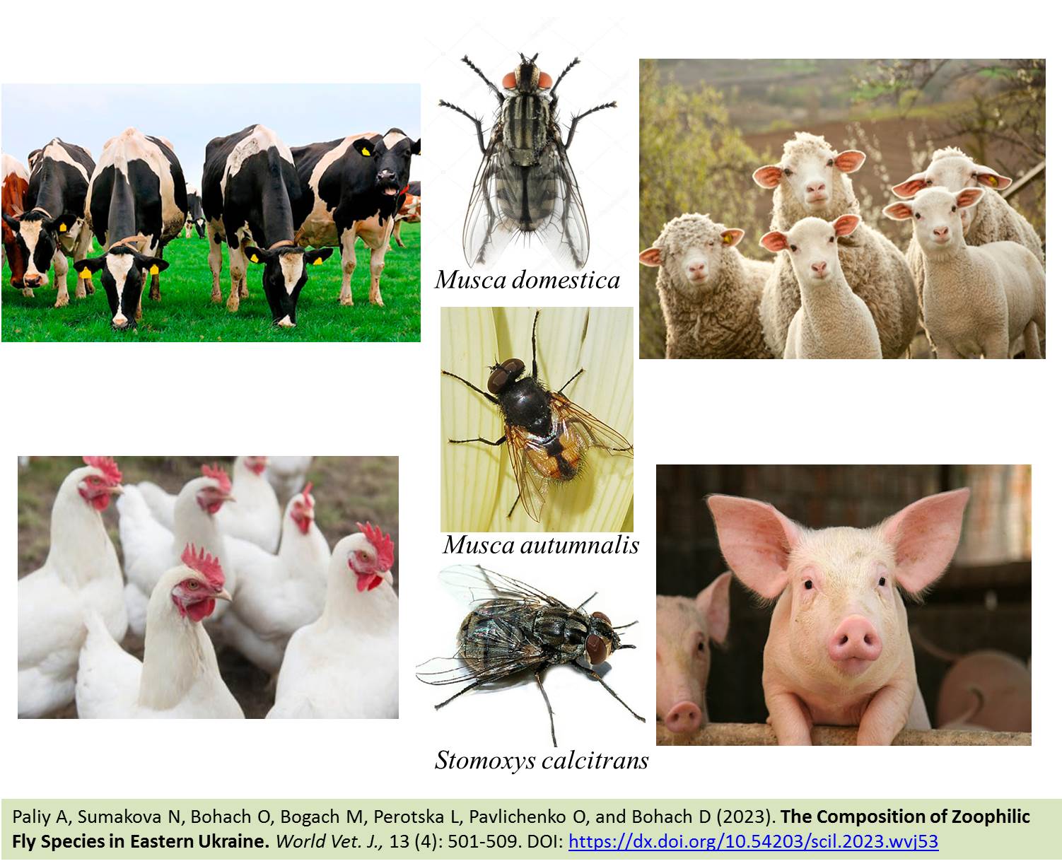
The Composition of Zoophilic Fly Species in Eastern Ukraine
Paliy A, Sumakova N, Bohach O, Bogach M, Perotska L, Pavlichenko O, and Bohach D.
World Vet. J. 13(4): 501-509, 2023; pii:S232245682300053-13
DOI: https://dx.doi.org/10.54203/scil.2023.wvj53
ABSTRACT: Zoophilic Diptera plays a leading role in the epizootic foci formation of many infectious and parasitic diseases and directly affects the quality of livestock products. The current study aimed to analyze the number and species composition of parasitic Diptera in industrial, farm, and homestead agrobiocenoses of large and small cattle, pig, and poultry farms in Eastern Ukraine. The research involved entomological collection during the peak activity daylight hours in early May, July, and early September 2021-2022 per farm. A total of 360 entomological collections were made, and 4310 zoophilous flies were examined. In livestock farms of five districts of the Kharkiv region of Ukraine, 28 species of zoophilic flies were registered, among which Musca domestica, Muscina stabulans, Stomoxys calcitrans, Lucilia sericata, Protophormia terraenovae, and Drosophila species were dominant species. The analysis revealed that cattle biocenoses hosted 27 fly species, pigs had 8 species, and poultry and small cattle each had 7 species. The study indicated an increase in the population of Musca autumnalis, the main species in the pastures, near livestock premises during the summer. Stomoxys calcitrans was also recorded in livestock agrobiocenoses. The species Musca domestica, Musca autumnalis, and Stomoxys calcitrans account for 78.8% to 88.3% of the entire complex of zoophilous flies. The two species of Ortellia caesarion (shiny dung beetle) and Ortellia cornicina (green dung beetle), known for their role as manure mineralizers and deemed non-threatening to animals, were completely absent during the research period. The findings indicated the species of Eristalis tenax in agrobiocenoses in 2021. Therefore, it can be concluded that zoophilic flies are physical irritants to animals and potential carriers of many infectious diseases, especially diseases caused by unicellular organisms.
Keywords: Biotopes, Musca autumnalis, Musca domestica, Stomoxys calcitrans, Zoophilic flies
[Full text-PDF] [Crossref Metadata] [Scopus] [Export from ePrints]
 Research Paper
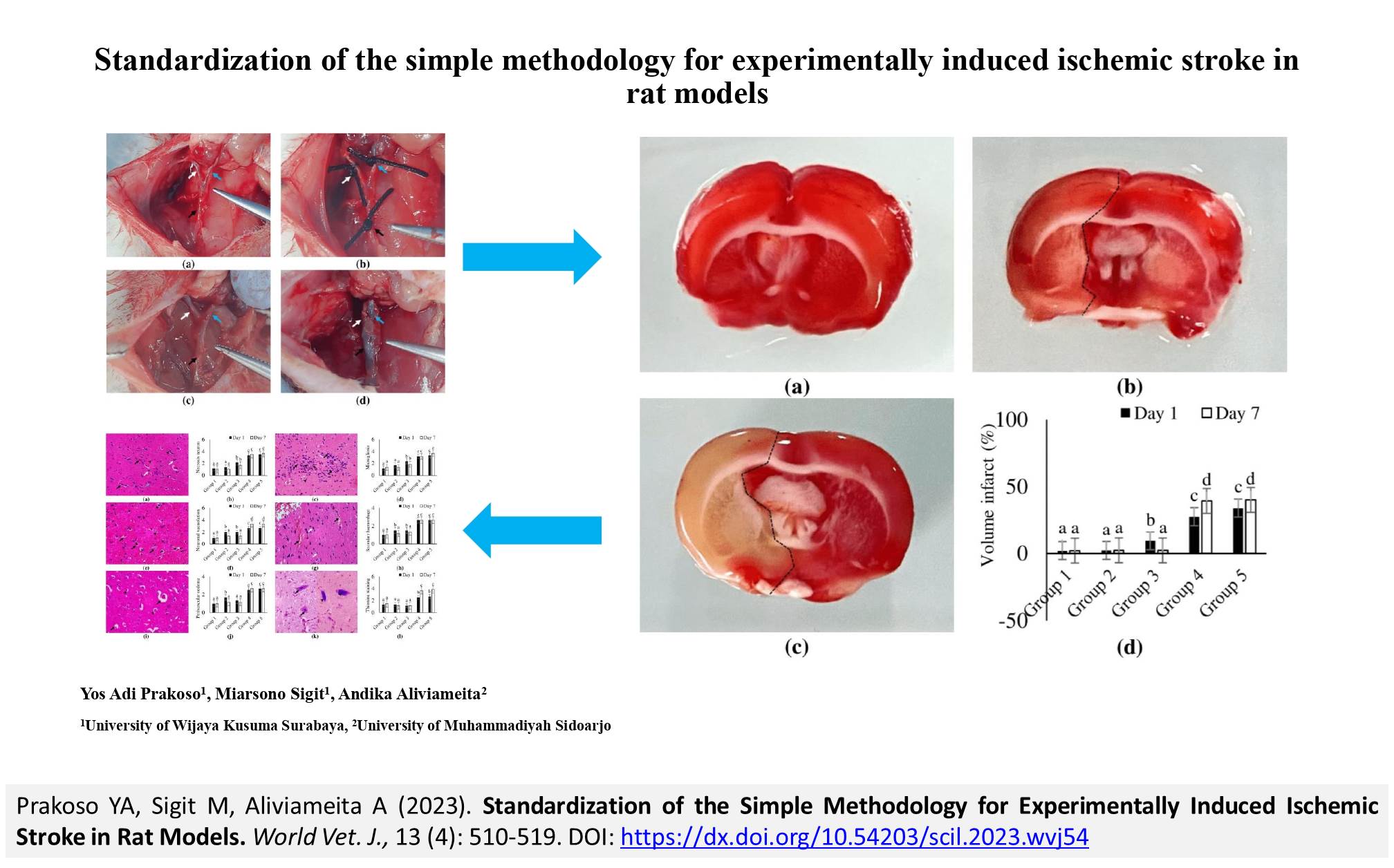
Standardization of the Simple Methodology for Experimentally Induced Ischemic Stroke in Rat Models
Prakoso YA, Sigit M, Aliviameita A.
World Vet. J. 13(4): 510-519, 2023; pii:S232245682300054-13
DOI: https://dx.doi.org/10.54203/scil.2023.wvj54
ABSTRACT: Stroke is a globally significant and devastating disease that requires prompt treatment. Animal models are commonly used to investigate stroke therapy, often through experimentally induced ischemic stroke (EIIS). However, challenges arise in implementing EIIS in animal models. The current study aimed to present a simple EIIS methodology for animal models. A total of 60 male Sprague-Dawley rats were randomly divided into five groups, namely Group 1 (sham-operated), Groups 2 to 5 (EIIS groups) with different duration of common carotid artery (CCA) ligation, including 1, 2, 4, and 8 hours, respectively. The ligation was performed on the CCA and its branches. Before the experiment, the rats were anesthetized, and the incision area was shaved and disinfected. The sagittal ventral midline was incised, with neck muscles retracted to expose the right CCA. The occlusion was performed on three sides of a carotid artery (common, external, and internal) using a simple interrupted suture. The occlusion of blood flow using ligation was performed at different times depending on the groups. After that, the CCA ligations were re-perfused by cutting the suture knot. The brain and blood were collected on days 1 and 7 after reperfusion. The results indicated that 4 and 8 hours of CCA ligation significantly impacted the general condition and neuro-deficit score. Moreover, 4 and 8 hours of CCA ligation could induce ischemic stroke by its capacity to cause infarction within the brain parenchyma and increase the platelet-to-white blood cell ratio, C-reactive protein, and De Ritis ratio. In contrast, 1 and 2 hours of CCA ligation did not significantly affect the observed parameters. It can be concluded that the EIIS using 4 and 8 hours of CCA ligation can be applied to induce ischemic stroke in rat models with consistent impacts on general conditions, neuro-deficit, hematology, and serology.
Keywords: Common carotid artery, Ischemic stroke, Ligation, Rat model, Standardization
[Full text-PDF] [Crossref Metadata] [Scopus] [Export from ePrints]
 Research Paper
Carrageenan-Induced Acute Inflammation on Back-Skin of Mice: Histopathological Features, Number of Inflammatory Cells, and Expression of COX-2, COX-1, and IL-6
Widyarini S, Sugiyono, Akrom AM, and Paryuni AD.
World Vet. J. 13(4): 520-530, 2023; pii:S232245682300055-13
DOI: https://dx.doi.org/10.54203/scil.2023.wvj55
ABSTRACT: Carrageenan is a sulfated polysaccharide obtained from red seaweed (Rhodophyceae) and can trigger inflammatory activation in both humans and laboratory animals. This study aimed to investigate the expression of cyclooxygenase-2 (COX-2), cyclooxygenase-1 (COX-1), and interleukin-6 (IL-6) and the number of inflammatory cells (neutrophil) involved in a carrageenan-induced acute inflammatory model in the back skin of mice. Paraffin blocks from the back skin of female Swiss mice aged 8 weeks were used in this study. The back-skins of 4 groups of 5 mice in each group were subcutaneously injected with 1%, 2%, and 4% carrageenan powder in 0.9% buffer saline and 0.9% buffer saline as control. Skin samples on paraffin blocks were taken 6 hours after carrageenan injection. Furthermore, paraffin blocks were stained with hematoxylin-eosin (HE) to count the number of inflammatory cells. Immunohistochemistry staining using anti-COX-2, COX-1, and IL-6 antibodies was performed to determine the role of inflammatory mediators. The results showed that the number of inflammatory cells (neutrophils) increased significantly following an increase in carrageenan concentrations. The COX-2, COX-1, and IL-6 expressed by inflammatory cells increased significantly at carrageenan concentrations of 1% to 4%. Histopathological features supported the results obtained from the calculation of the number of inflammatory cells and the expression of COX-2, COX-1, and IL-6. The inflammatory markers consisting of COX-2, COX-1, and IL-6 were expressed on the back skin of mice at 6 hours post-injection with 1% to 4% carrageenan. It can be concluded that carrageenan can be used for an acute inflammatory model of the back skin of a mouse. This inflammation model is intended to facilitate the evaluation or measurement of therapeutic and inflammatory responses when test substances are administered topically or transdermal.
Keywords: Carrageenan, Cyclooxygenase-2, Cyclooxygenase-1, Interleukin-6, Inflammatory cell, Skin inflammation
[Full text-PDF] [Crossref Metadata] [Scopus] [Export from ePrints]
 Research Paper
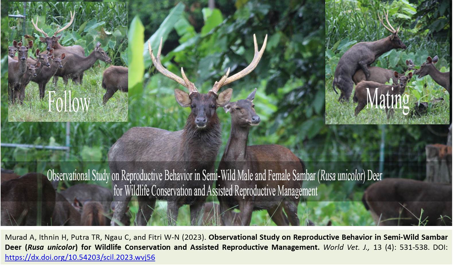
Observational Study on Reproductive Behavior in Semi-Wild Sambar Deer (Rusa unicolor) for Wildlife Conservation and Assisted Reproductive Management
Murad A, Ithnin H, Putra TR, Ngau C, and Fitri W-N.
World Vet. J. 13(4): 531-538, 2023; pii:S232245682300056-13
DOI: https://dx.doi.org/10.54203/scil.2023.wvj56
ABSTRACT: Understanding the reproductive behaviors of different wildlife species is essential to unravel their reproductive strategies, ecological adaptations, and conservation requirements. This study delved into the reproductive biology of the sambar deer (Rusa unicolor), with a focus on promoting assisted reproductive technology for wildlife conservation and investigating the reproductive behaviors of male and female sambar deer. The study was conducted at Pusat Konservasi Hidupan Liar (PKHL) Sungkai, Perak, Malaysia. The observation focused on one male and two female sambar deer. Direct observations of the deer were conducted for 14 days in September 2022. The direct observations were performed in the morning (Session 1= 8-10 am), afternoon (Session 2 = 10-12 pm), and evening (Session 3= 3-5 pm), using the instantaneous sampling method. A total of 75 behavior instances were recorded, in which male deer exhibited the most reproductive behavior at 58 instances (77.3% of the total reproductive behavior). Successful mating was observed on day 6, elucidating a crepuscular preference in the male animal in exhibiting reproductive behavior. The female’s reproductive behavior lasted for a short period, from 24 hours for Female 2 and 72 hours for Female 1. In conclusion, there was a distinct behavior between the male and female deer during the rutting season. Understanding the reproductive behavior to estimate the length of estrus can be useful as a non-invasive tool to detect heat and can be considered to improve breeding management and implement assisted reproductive technology.
Keywords: Breeding, Conservation, Ex-situ, Release program, Wildlife
[Full text-PDF] [Crossref Metadata] [Scopus] [Export from ePrints]
 Research Paper
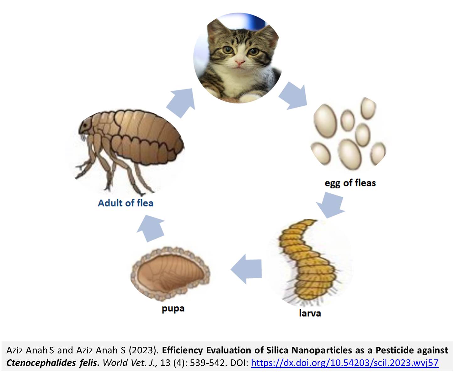
Efficiency Evaluation of Silica Nanoparticles as a Pesticide against Ctenocephalides felis
Aziz Anah S and Aziz Anah S.
World Vet. J. 13(4): 539-542, 2023; pii:S232245682300057-13
DOI: https://dx.doi.org/10.54203/scil.2023.wvj57
ABSTRACT: The increasing resistance of arthropods to many insecticides has encouraged researchers to search for new alternatives to combat harmful insects. The present study aimed to evaluate the effectiveness of silica nanoparticles (NPs) on Ctenocephalides felis (C. felis), a prevalent species among cats and a known vector for diseases. The killing efficacy of SiO2-NPs against C. felis was tested at three different concentrations (50, 100, and 150 mg/ml) over three different time intervals (10, 20, and 40 minutes), alongside positive and negative control groups (distilled water and cypermethrin). The results of the current study indicated that all concentrations had a fleacidal effect, with SiO2-NPs demonstrating increased efficacy with higher concentrations and longer exposure periods. The concentration of 150 mg/mL of SiO2-NPs led to the highest effect at 96% upon exposure for 40 minutes. The results of the current study revealed significant differences between the control groups and all the groups treated with Sio2-NP concentrations. It can be concluded that Sio2-NPs are a practical approach to flea control although it is necessary to search for environmentally friendly pesticides. The current results indicate that SiO2-NPs have anti-parasitic effects against C. felis.
Keywords: Cat fleas, Iraq, Nanoparticles, Pesticide, Silica
[Full text-PDF] [Crossref Metadata] [Scopus] [Export from ePrints]
 Research Paper
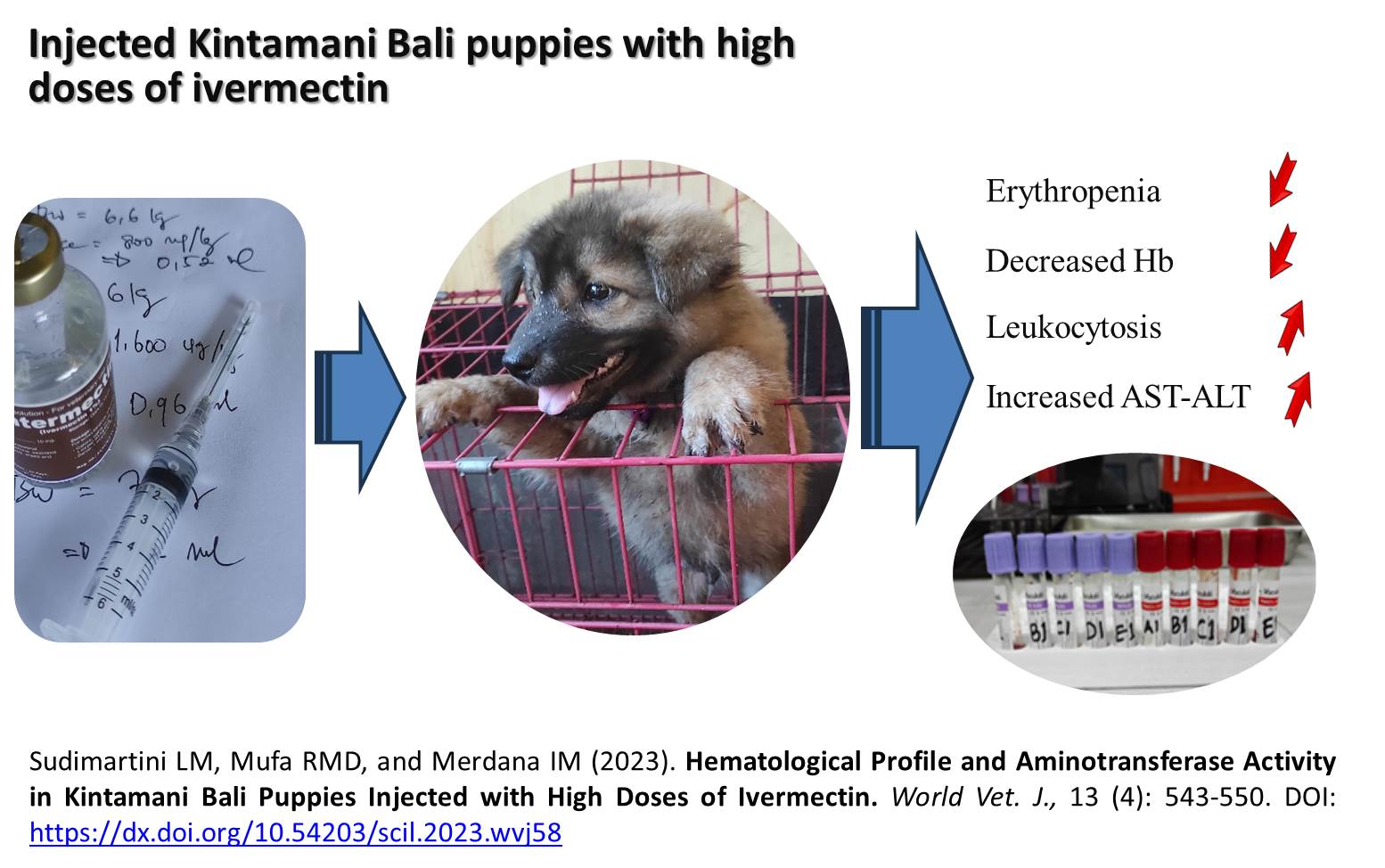
Hematological Profile and Aminotransferase Activity in Kintamani Bali Puppies Injected with High Doses of Ivermectin
Sudimartini LM, Mufa RMD, and Merdana IM.
World Vet. J. 13(4): 543-550, 2023; pii:S232245682300058-13
DOI: https://dx.doi.org/10.54203/scil.2023.wvj58
ABSTRACT: Ivermectin toxicity is known to cause harmful side effects or even death in dogs intolerant to the medication. Intolerant dogs have a mutation in the MDR-1 (Multi-Drug Resistance) gene, so they lack the P-glycoprotein gene that removes drugs from the brain. Therefore, this study aimed to determine ivermectin toxicity in Kintamani Bali puppies by examining physiological responses based on hematological profiles and aminotransferase activity after a high-dose injection. A laboratory observational approach was used, and the samples were 25 healthy female Kintamani puppies based on a veterinary examination, aged 3-6 months, weighing 6.32 ± 1.18 kg, randomly divided equally into five treatment groups. The treatments included a placebo (1ml Aqua Pro Injection) as a control, as well as a single dose of ivermectin injection sequentially 200, 400, 800, and 1600 µg/kg subcutaneously. Blood samples were collected before treatment and after 7 and 14 days post-treatment. The hematologic parameters observed included levels of hemoglobin, erythrocytes, hematocrit, total leukocytes, neutrophils, lymphocytes, monocytes, eosinophils, and basophils, as well as blood biochemistry, namely aspartate aminotransferase (AST) and alanine aminotransferase (ALT) activities. Observation results after 4 hours of administration of ivermectin at doses of 800 and 1600 µg/kg of puppies showed changes in behavior, restlessness, depression, tremors, mydriasis, hypersalivation, anorexia, and polydipsia. Meanwhile, the results of hematological examination on the seventh day after ivermectin treatment showed a trend of erythropenia, leukocytosis, a decrease in hemoglobin levels, and an increase in aminotransferase enzyme activity. This condition continued until day 14, but the physiological parameter values showed that the puppy’s condition gradually improved compared to the seventh day after treatment. There were significant differences in the blood profile, AST, and ALT of Kintamani puppies injected with ivermectin at doses of 800 and 1,600 ug/kg compared to controls on days 7 and 14 after and before treatment. It was concluded that high-dose ivermectin injections in Kintamani Bali puppies caused toxicity with clinical signs of erythropenia, decreased hemoglobin, leukocytosis, and increased aminotransferase activity.
Keywords: Aminotransferase, Blood profile, Ivermectin, Kintamani dogs, Toxicity
[Full text-PDF] [Crossref Metadata] [Scopus] [Export from ePrints]
Research Paper
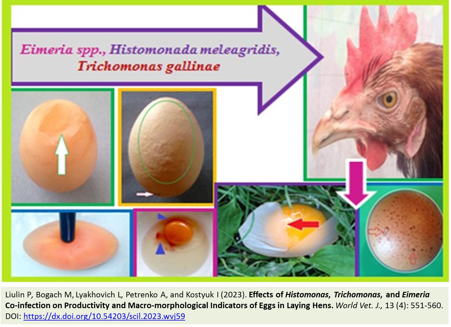
Effects of Histomonas, Trichomonas, and Eimeria Co-infection on Productivity and Macro-morphological Indicators of Eggs in Laying Hens
Liulin P, Bogach M, Lyakhovich L, Petrenko A, and Kostyuk I.
World Vet. J. 13(4): 551-560, 2023; pii:S232245682300059-13
DOI: https://dx.doi.org/10.54203/scil.2023.wvj59
ABSTRACT: The study of macro-morphological changes is important for recognizing disturbances in egg formation that cause pathologies, especially co-infection. The current study aimed to evaluate the level of egg productivity and macro-morphological parameters of eggs in domestic chickens of the Rhode Island breed with co-infection of Histomonas, Trichomonas, and Eimeria. Clinical and parasitological, coproscopic, morphometric research, and statistical analysis methods were used for this research. Pathogens of Histomonas and Trichomonas were detected by microscopy of smears of fresh feces, and Eimeria oocysts were identified by flotation according to the Fullenborn method. During 30 days of research, there was a significant decrease in egg production (52%), a decrease in egg weight by 16.8%, and a decrease in the shell thickness by 30.43% during spontaneous Eimeria-Histomonosis-Trichomonosis co-infection in laying hens. The eggshell indicated noticeable macro-morphological changes, including deformations and defects resulting from insufficient calcification. These changes manifest as combined damage to the shell, characterized by small cracks, roughness, bumpy or spilled thickenings, and complete or partial depigmentation. When evaluating the internal content of eggs in 12% of their samples, there were bloody spots, relatively smaller and lighter yolks, thinning of the protein part. Thus, the specified macro-morphological changes and egg defects were the result of the negative impact of co-infection on the processes of egg formation, which indicates the systemic nature of the lesion and the morphofunctional insufficiency of the egg-forming organs.
Keywords: Comorbidity, Egg defect, Egg production, Eimeriosis, Histomonosis, Laying hen, Trichomoniasis
[Full text-PDF] [Crossref Metadata] [Scopus] [Export from ePrints]
 Research Paper
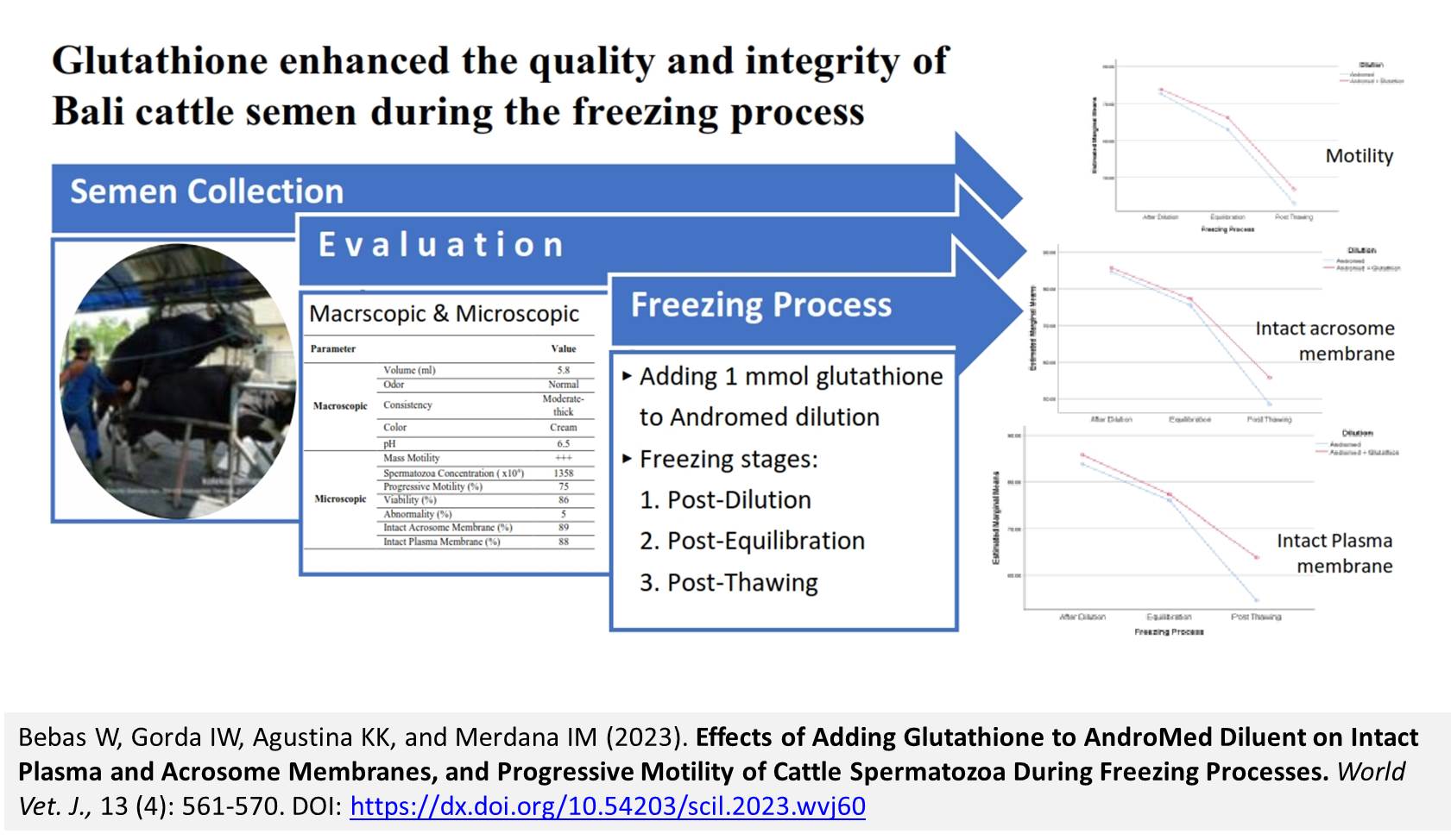
Effects of Adding Glutathione to AndroMed Diluent on Intact Plasma and Acrosome Membranes, and Progressive Motility of Cattle Spermatozoa During Freezing Processes
Bebas W, Gorda IW, Agustina KK, and Merdana IM.
World Vet. J. 13(4): 561-570, 2023; pii:S232245682300060-13
DOI: https://dx.doi.org/10.54203/scil.2023.wvj60
ABSTRACT: Adding endogenous antioxidants to the diluent is significantly associated with semen quality during the freezing process. This study aimed to investigate the effects of adding glutathione to AndroMed diluent on the preservation of crucial sperm attributes, namely, intact plasma membrane (IPM), intact acrosome membrane (IAM), and progressive motility of Bali cattle spermatozoa. A completely randomized design was used, and spermatozoa samples were obtained from a Bali cattle and divided into two diluent treatment groups (36 diluent samples in each group with six replications), namely pure AndroMed as the control and a group with the addition of glutathione (1 mmol) to AndroMed. Each treatment was replicated six times and evaluated at three freezing stages, including post-dilution, post-equilibration, and post-thawing, for crucial sperm properties. The results indicated that fresh Bali cattle spermatozoa had progressive motility, IAM, and IPM of 75%, 89%, and 88%, respectively. During the freezing process, there was a significant decrease in semen quality, including progressive motility, IAM, and IPM of spermatozoa after dilution to post-equilibration and post-equilibration to post-thawing in both treatment groups. Meanwhile, the addition of 1 mmol of glutathione to AndroMed diluent had a significant difference in increasing progressive motility, IAM, and IPM of Bali cattle spermatozoa at each stage of semen freezing, including post-dilution, post-equilibration, and post thawing when compared with controls. Based on the results, it can be concluded that adding 1 mmol of glutathione to the AndroMed diluent enhanced the quality and integrity of Bali cattle semen, including progressive motility, IAM, and IPM during the freezing process.
Keywords: Bali cattle, Freezing Process, Glutathione, Progressive Motility
[Full text-PDF] [Crossref Metadata] [Scopus] [Export from ePrints]
 Research Paper
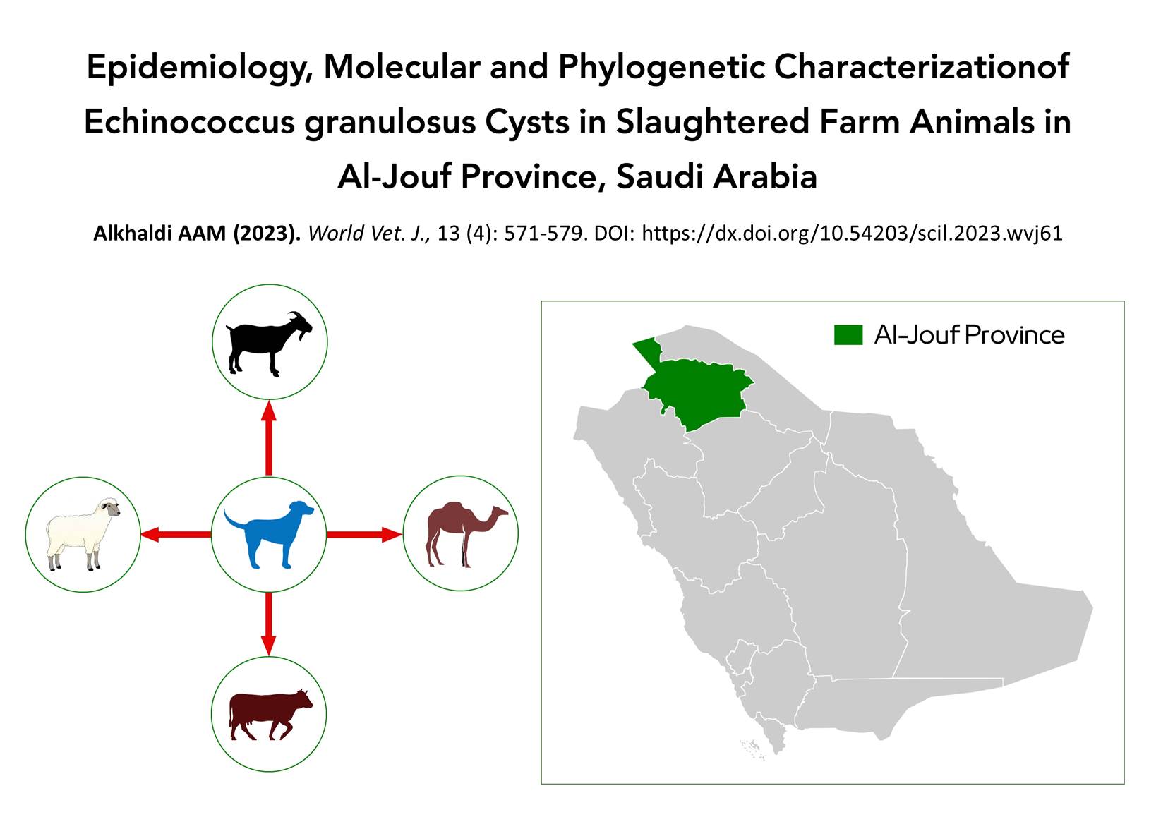
Epidemiology, Molecular, and Phylogenetic Characterization of Echinococcus granulosus Cysts in Slaughtered Farm Animals in Al-Jouf Province, Saudi Arabia
Alkhaldi AAM
World Vet. J. 13(4): 571-579, 2023; pii:S232245682300061-13
DOI: https://dx.doi.org/10.54203/scil.2023.wvj61
ABSTRACT: Echinococcosis, or hydatidosis, is a serious veterinary disease and public health issue worldwide, particularly in rural areas in which dogs have frequent contact with local herbivores. This study assessed the frequency of hydatidosis found among farm animals slaughtered in Al-Jouf Province in northern Saudi Arabia in 2021. A total of 156754 sheep, 36337 goats, 8590 camels, and 986 cattle were inspected for hydatidosis infection by comprehensive evaluation involving meticulous visual inspection and manual exploration of the internal organs through palpation. The cysts were subjected to molecular and phylogenetic analysis. The overall prevalence rates of hydatid cysts were 0.43%, 0.19%, 0.54%, and 0.51% in the inspected sheep, goats, camels, and cattle, respectively. The highest disease prevalence rates among sheep (27.8%) and goats (30.9%) occurred in the spring, and the highest prevalence rates among camels (41.3%) and cattle (80%) were in the summer. Regarding the prevalence of the disease in four slaughterhouses in the Al-Jouf Province, the highest prevalence in sheep, goats, and camels was in the Tabarjal slaughterhouse (1.43%, 0.81%, and 1.08%, respectively), although the Al-Qurayat slaughterhouse had the highest prevalence rate in cattle (1.98%). Complete molecular analysis indicated that cytochrome c oxidase subunit 1 (cox1) sequences from cyst isolates belonged to Echinococcus granulosus (E. granulosus). Moreover, there was high homology (98-100%) with associated Genbank sequences of E. granulosus isolated from sheep in the Kingdom of Saudi Arabia (KSA). Sheep and camels were a significant source of hydatidosis transmission to dogs and helped to maintain disease incidence in the Al-Jouf Province. Thus, significant efforts should focus on preventing cyst transmission from abattoirs and infected stray dogs.
Keywords: Echinococcus granulosus, Epidemiology, Molecular characterization, Farm animals
[Full text-PDF] [Crossref Metadata] [Scopus] [Export from ePrints]
 Research Paper
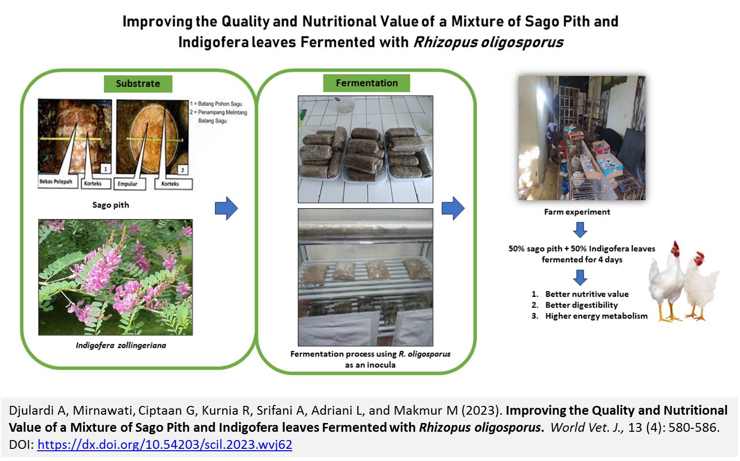
Improving the Quality and Nutritional Value of a Mixture of Sago Pith and Indigofera leaves Fermented with Rhizopus oligosporus
Djulardi A, Mirnawati, Ciptaan G, Kurnia R, Srifani A, Adriani L, and Makmur M.
World Vet. J. 13(4): 580-586, 2023; pii:S232245682300062-13
DOI: https://dx.doi.org/10.54203/scil.2023.wvj62
ABSTRACT: The nutritional value of sago pith is limited due to its low protein content, making it less suitable for poultry feed. To increase the benefit values of the sago pith, it is necessary to process it through fermentation. The current study aimed to determine the effects of substrate composition and fermentation time of fermented sago (Metroxylon sagu) pith (SP) and Indigofera (Indigofera zollingeriana) leaves (IL) mixture using Rhizopus oligosporus as an inoculum on crude protein, crude fat and crude fiber content of fermented SP and IL, nitrogen retention, crude fiber digestibility, and energy metabolism in broiler chickens. The study was performed on 30 broiler chickens, average weighing ± 1.5 kg at 6 weeks of age, along with SP, IL, and R. oligosporus. This experiment was conducted using a randomized design in a 3×3 factorial with three replications. Substrate composition, or factor A, was made up of A1 (80% SP + 20% IL), A2 (60% SP + 40% IL), and A3 (50% SP + 50% IL). Fermentation time as Factor B entailed B1 (2 days), B2 (3 days), and B3 (4 days). The findings demonstrated a significant interaction between the time of fermentation and the composition of the substrate in relation to crude protein content, nitrogen retention, crude fat, crude fiber digestibility, and energy metabolism. It can be concluded that the composition of substrate 50% SP and 50% IL with 3 days of fermentation yielded the best result, with crude protein at 25.45%, nitrogen retention at 59.72%, crude fat at 0.020%, crude fiber at 6.40%, crude fiber digestibility at 57.34%, and metabolic energy at 2658.44 kcal/kg.
Keywords: Broiler chickens, Crude protein, Indigofera, Sago pith, Rhizopus oligosporus
[Full text-PDF] [Crossref Metadata] [Scopus] [Export from ePrints]
 Research Paper
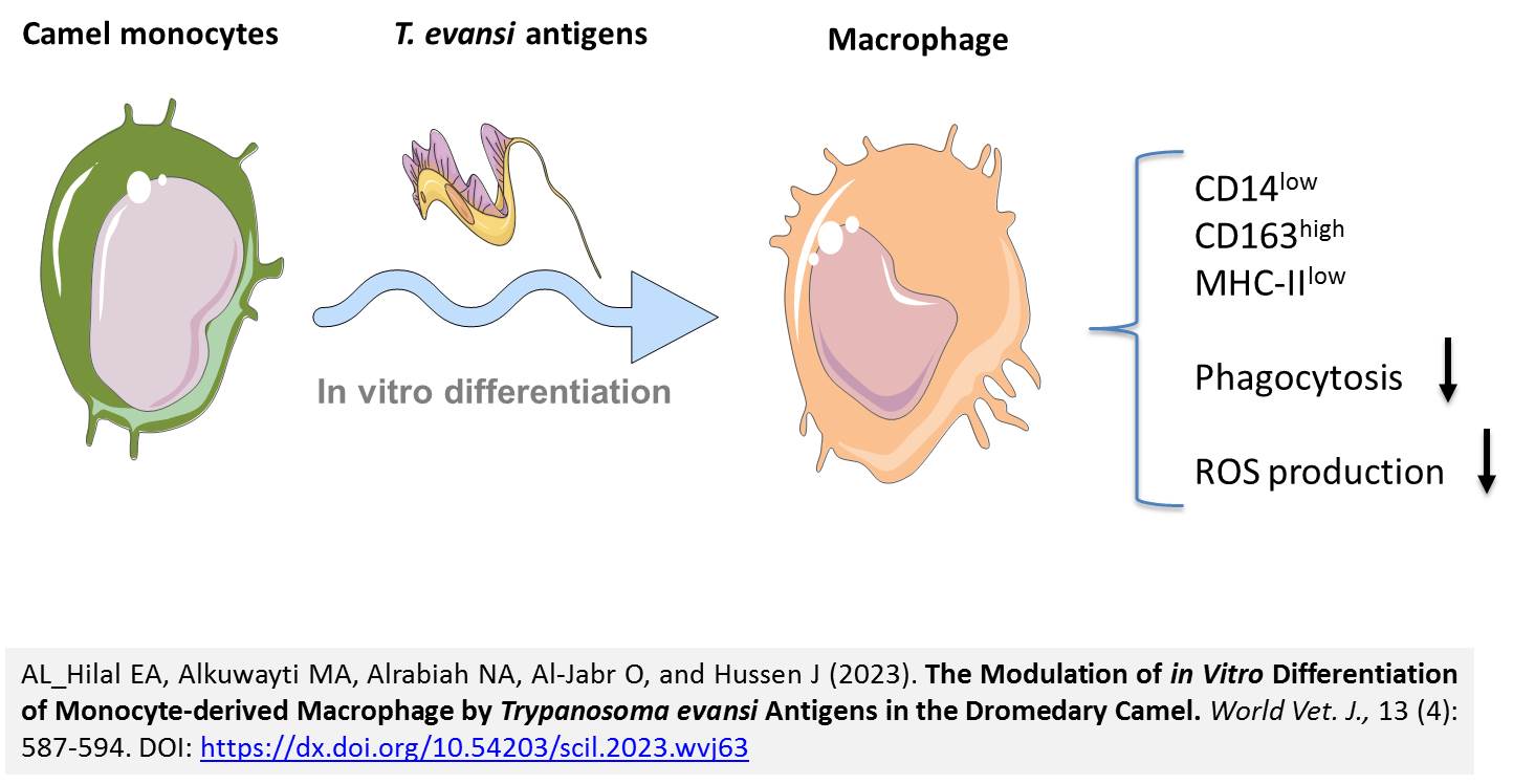
The Modulation of in Vitro Differentiation of Monocyte-derived Macrophage by Trypanosoma evansi Antigens in the Dromedary Camel
AL_Hilal EA, Alkuwayti MA, Alrabiah NA, Al-Jabr O, and Hussen J.
World Vet. J. 13(4): 587-594, 2023; pii:S232245682300063-13
DOI: https://dx.doi.org/10.54203/scil.2023.wvj63
ABSTRACT: Studies on the camel immune response to Trypanosoma (T.) evansi, the causative agent of Surra, are very limited. In the present study, flow cytometry was employed to investigate the modulatory effects of different T. evansi antigens on the in vitro differentiation of camel blood monocytes into macrophages. For this, in vitro, separated camel monocytes were differentiated into monocyte-derived macrophages (MDM) in the presence or absence (control) of formalin-fixed (inactivated) T. evansi whole parasite (T. evansi group) or the purified Ro Tat 1.2 antigen (Ro Tat 1.2 group). The analysis of the antimicrobial functions of MDM (phagocytosis and reactive oxygen species (ROS) production) revealed reduced phagocytosis activity of camel MDM generated in the presence of T. evansi antigens. In addition, a lack of ROS-response was observed in camel MDM generated in the presence of T. evansi antigens after stimulation with PMA. These results indicated a compromising effect of T. evansi on the innate defense mechanisms in camels. Phenotypic analysis revealed the upregulation of major histocompatibility complex (MHC) class II molecules together with the lower abundance of the scavenger receptor for haptoglobin–hemoglobin complexes (CD163) on MDM generated in the presence of whole T. evansi parasites, indicating a polarizing effect of T. evansi on the differentiation of camel monocytes into an M1 phenotype. However, the reduced antimicrobial functions of these cells argue against their pro-inflammatory nature. Although both MDM generated in the presence of whole T. evansi antigens or their purified Ro Tat 1.2 proteins indicated similar expression levels of CD14 and MHCII molecules, the different abundance of the cell surface molecules CD172a, CD163, CD45, and CD44 indicated different phenotypes of the two MDMs. The results of the present study revealed compromising effects of T. evansi antigens on camel macrophages differentiated in vitro from blood monocytes. Whether these effects contribute to the in vivo pathogenesis of T. evansi in camels remains to be determined in future studies.
Keywords: Camel, Flow cytometry, Immunity, Macrophage, Monocyte, Trypanosoma evansi
[Full text-PDF] [Crossref Metadata] [Scopus] [Export from ePrints]
 Review
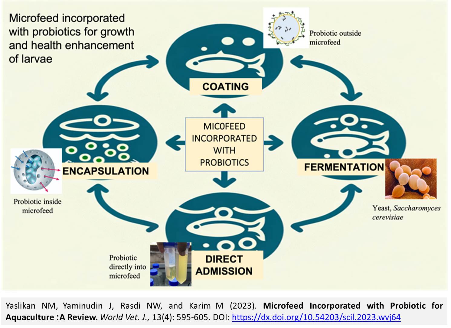
Microfeed Incorporated with Probiotic for Aquaculture: A Review
Yaslikan NM, Yaminudin J, Rasdi NW, and Karim M.
World Vet. J. 13(4): 595-605, 2023; pii:S232245682300064-13
DOI: https://dx.doi.org/10.54203/scil.2023.wvj64
ABSTRACT: Ensuring the availability of high-quality larvae in sufficient quantities remains a significant bottleneck for the grow-out phase of aquaculture. Over the past century, various alternative dietary solutions for larval stages have been explored, encompassing bacteria, microalgal pastes, yeasts, and various inert microparticles, though with inconsistent outcomes. This review aimed to discuss the innovative integration of probiotics into microfeeds, highlighting encapsulation, coating, and fermentation techniques to propel aquaculture productivity. Microfeeds, which are often nutrient-rich and easily assimilated in powdered or liquid form, play a crucial role in larval fish nutrition. These can be classified into microencapsulated, dry, liquid, and live feeds. The choice of microfeed is pivotal, ensuring appeal, digestibility, and water stability tailored to each larval stage. As probiotics gain popularity in aquaculture for their potential to enhance growth, bolster disease resistance, and improve water quality, their administration methods have diversified. The probiotics can be administered through direct immersion and bath treatments to biofloc systems and feed additives. The results indicated that microfeed incorporated with probiotics showed a positive result impact on the aquaculture industry.
Keywords: Alternative diets, Aquaculture, Microfeed, Probiotics
[Full text-PDF] [Crossref Metadata] [Scopus] [Export from ePrints]
 Review
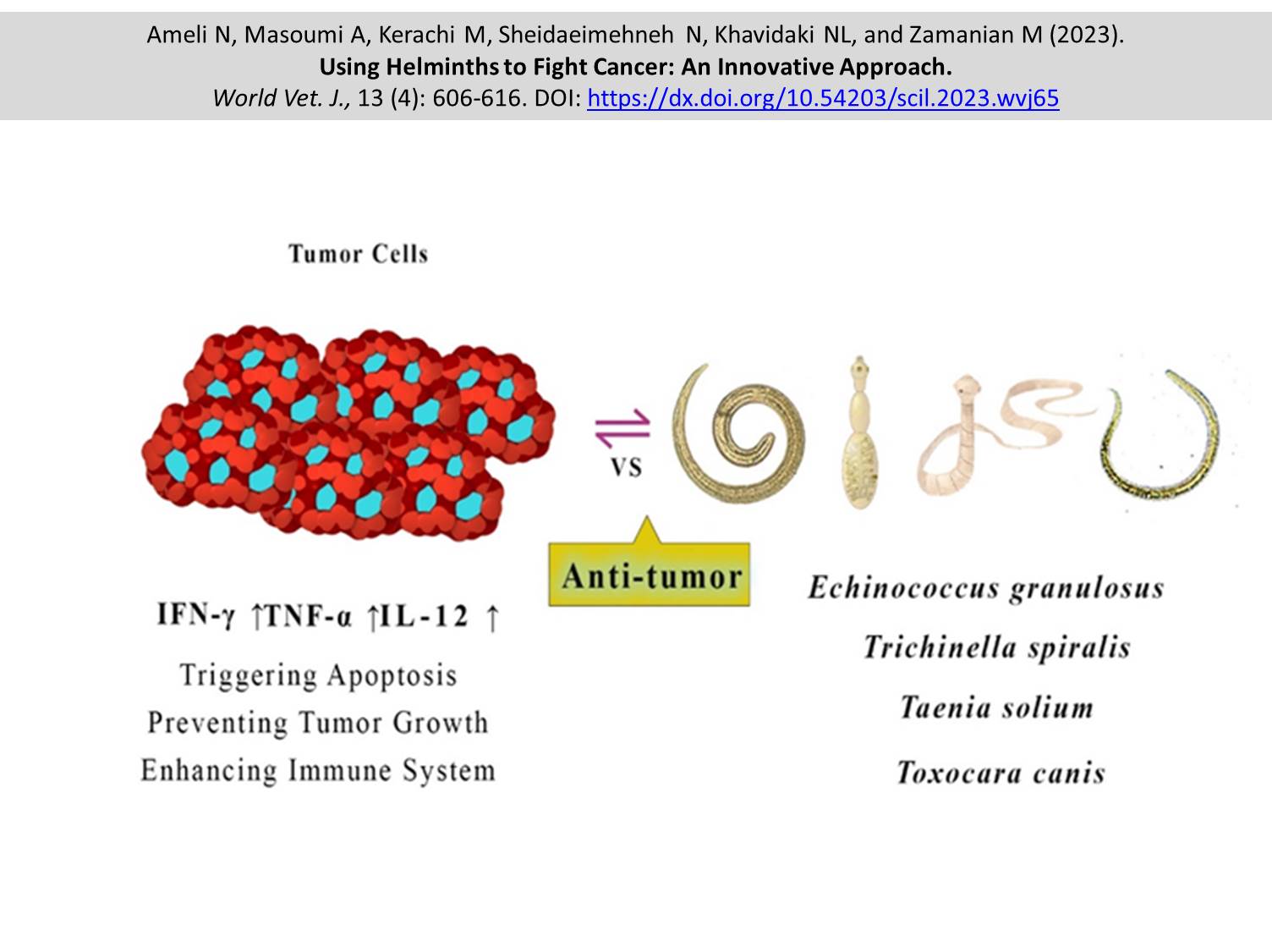
Using Helminths to Fight Cancer: An Innovative Approach
Ameli N, Masoumi A, Kerachi M, Sheidaeimehneh N, Khavidaki NL, and Zamanian M.
World Vet. J. 13(4): 606-616, 2023; pii:S232245682300065-13
DOI: https://dx.doi.org/10.54203/scil.2023.wvj65
ABSTRACT: As an alternative treatment in cancer therapy, there has been a growing interest in using helminths, such as Trichinella spiralis (T. spiralis), Echinococcus granulosus (E. granulosus), Toxocara canis (T. canis), and Taenia solium (T. solium). This study aimed to investigate the antigens and mechanisms that contribute to the anticancer properties of helminths, providing insights into how helminths may be used as a new and innovative treatment modality for cancer. The current review analyzed preclinical and clinical studies published between 2000 and 2023. The present study sought to obtain information on helminths, such as E. granulosus, T. spiralis, T. canis, and T. solium, to treat cancers of the breast, pancreas, melanoma, and leukemia by exploring databases, such as PubMed, Google Scholar, and Scopus. Studies focusing on helminth therapy against particular cancer types for in vitro and animal models were included. Several studies have shown the possibility of inhibiting breast, colon, melanoma, and leukemia tumor growth, inducing apoptosis, and modulating the tumor microenvironment with E. granulosus, T. spiralis, T. canis, and T. solium based on in vitro and animal models studies. Some studies have indicated that helminth therapy can improve survival rates, reduce tumor growth, and stimulate the immune system in cancer patients. A potential improvement in treatment outcomes can be used for combination therapies, such as antigen selection, immune profiling, and individualized approaches based on helminth therapy. Helminth therapy is an additional option for cancer treatment, emphasizing T. spiralis, E. granulosus, T. canis, and T. solium. These helminth antigens could modulate immune responses and directly cause cytotoxicity in cancer cells.
Keywords: Cancer, Echinococcus granulosus, Taenia solium, Toxocara canis, Trichinella spiralis
[Full text-PDF] [Crossref Metadata] [Scopus] [Export from ePrints]
 Review
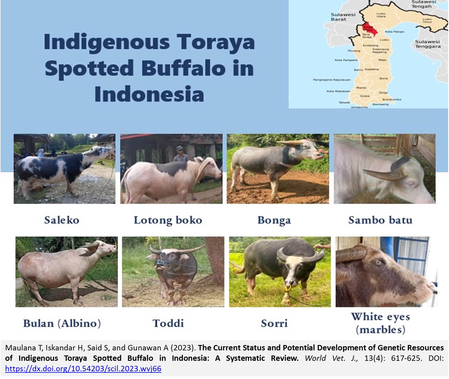
The Current Status and Potential Development of Genetic Resources of Indigenous Toraya Spotted Buffalo in Indonesia: A Systematic Review
Maulana T, Iskandar H, Said S, and Gunawan A.
World Vet. J. 13(4): 617-625, 2023; pii:S232245682300066-13
DOI: https://dx.doi.org/10.54203/scil.2023.wvj66
ABSTRACT: Buffaloes are integral to the Asiatic market as they are crucial for agricultural work and transportation and provide a significant source of dairy and meat, contributing to various industries, local economies, and cultural practices across the region. Indonesia is a mega biodiversity country abundant in livestock genetic resources, including indigenous, local, and introduced breeds that play a crucial role in agriculture and the livestock industry. These genetic resources offer the potential for selective breeding and improving the quality of livestock populations through well-designed breeding programs, ensuring sustainable livestock production for the future. The current study was performed using the “Publish or Perish” software, and the data obtained was analyzed using the CADIMA web tool. The Toraya buffalo population is the largest in the Tana Toraja and North Toraja regencies and is widely distributed within South Sulawesi Province, Indonesia. The population of Toraya buffalo in this region reached 43674 heads. Toraya buffaloes exhibit diverse body color characteristics, such as black, gray, white, and mixed. Moreover, Toraya buffaloes possess distinct quantitative traits that set them apart from other swamp buffalo breeds. Although there are limited studies on Toraya buffaloes, the potential for broader and more comprehensive studies offers opportunities to uncover new information on the characteristics, genetics, reproduction, health, and management of Toraya buffaloes. The development of Toraya buffalo farms also holds significant economic promise, as it can lead to increased agricultural productivity and improved livelihoods for local communities by enhancing the quality by implementing well-planned breeding programs and leveraging reproductive technology, and genetics-based selection, growth, and productivity can be produced. To increase the population and productivity of Toraya buffaloes, a well-structured breeding program integrating reproductive technology and selection based on quantitative and molecular genetics is essential. The development potential of Toraya buffalo is vast, not only due to its high cultural value but also its superior quantitative traits compared to common swamp buffalo, positioning it as a potential national meat provider. The present review article aimed to discuss the characteristics and development potential of Toraya buffaloes, along with the implementation of reproductive biotechnology and molecular genetics to enhance the population, productivity, and quality of Toraya buffaloes in Indonesia.
Keywords: Genetic resource, Spotted buffalo, Swamp buffalo, Toraya buffalo
[Full text-PDF] [Crossref Metadata] [Scopus] [Export from ePrints]
 Case Report
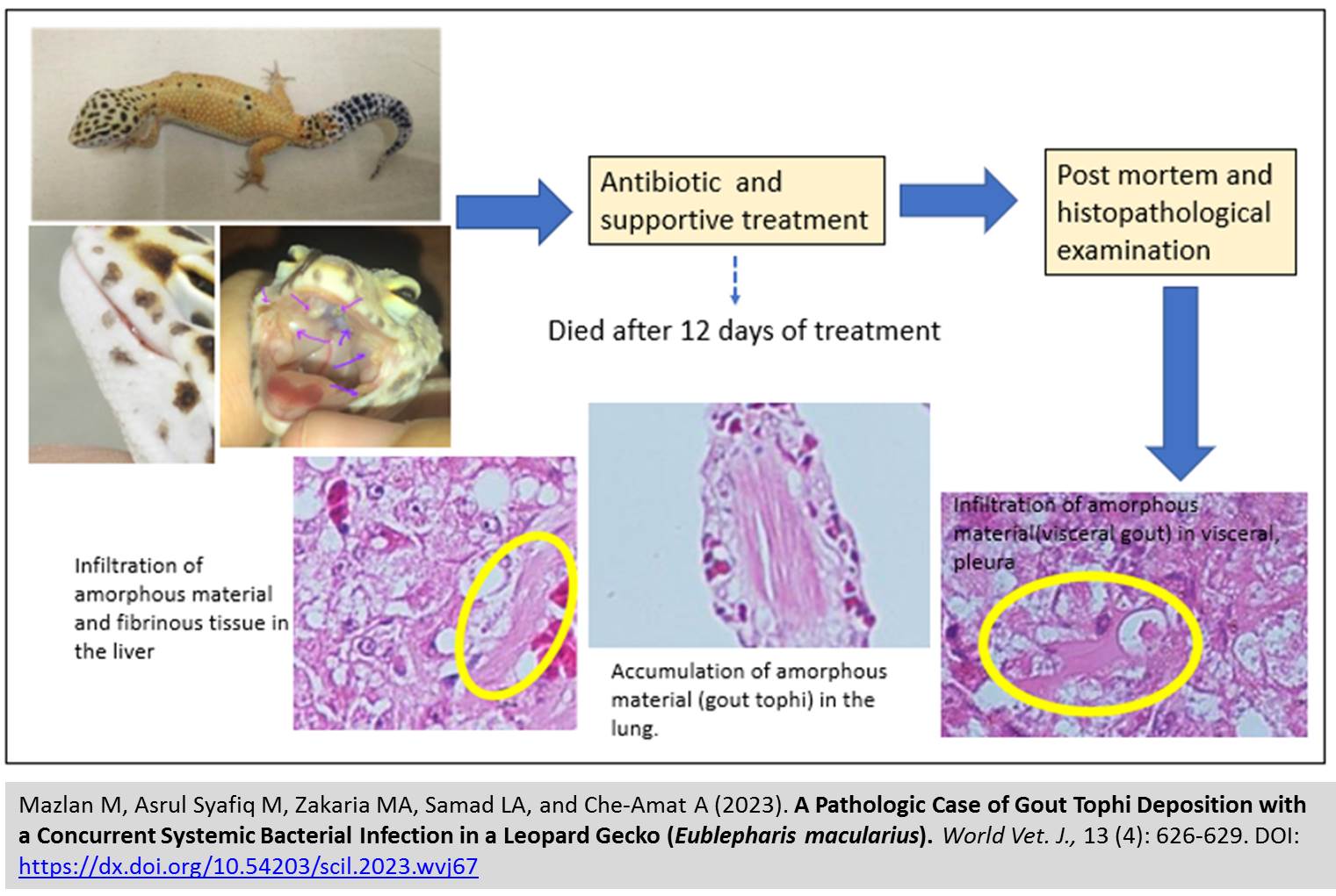
Pathologic Case of Gout Tophi Deposition with a Concurrent Systemic Bacterial Infection in a Leopard Gecko (Eublepharis macularius)
Mazlan M, Asrul Syafiq M, Zakaria MA, Samad LA, and Che-Amat A.
World Vet. J. 13(4): 626-629, 2023; pii:S232245682300067-13
DOI: https://dx.doi.org/10.54203/scil.2023.wvj67
ABSTRACT: Gout is caused by excessive uric acid in the blood deposited in tissues (visceral gout) or joints (articular gout), leading to severe inflammation and pain. A female leopard gecko was presented to the University Veterinary Hospital, University of Putra, Malaysia, with a history of swelling at the left caudal mandible, inappetence, and weight loss. An oral examination indicated a swollen mouth with scabs on the upper right mandible and multiple whitish deposits inside the mouth. The preliminary diagnosis was mouth rot, and the treatment included metronidazole and a multivitamin supplement. The leopard gecko died 12 days after treatment since there was no improvement. Post-mortem examination revealed that the liver was slightly enlarged with generalized moderate congestion and the presence of whitish deposits, as well as noticeable whitish deposits on the pleural surface of the lungs. Histopathological examination of the lungs revealed a granuloma with an inflammatory reaction predominantly by abundant mononuclear cells and fibrin deposition. An irregular collection of amorphous materials in the visceral pleura suggested gout tophi. The liver was infiltrated with amorphous material and fibrinous tissue, and it had mild congestion, indicating visceral gout and bacterial infection. Klebsiella pneumoniae and Proteus mirabilis were isolated from the lungs and liver samples, respectively. In conclusion, gout tophi is common in reptiles, but visceral involvement is rare, and early detection is critical to avoid secondary bacterial infection, as demonstrated in this case.
Keywords: Amorphous material, Bacterial infection, Gout tophi, Histopathology, Leopard gecko
[Full text-PDF] [Crossref Metadata] [Scopus] [Export from ePrints]
 Case Report
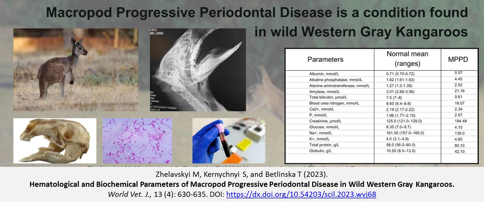
Hematological and Biochemical Parameters of Macropod Progressive Periodontal Disease in Wild Western Gray Kangaroos
Zhelavskyi M, Kernychnyi S, and Betlinska T.
World Vet. J. 13(4): 630-635, 2023; pii:S232245682300068-13
DOI: https://dx.doi.org/10.54203/scil.2023.wvj68
ABSTRACT: Macropod progressive periodontal disease (MPPD), known as Lumpy Jaw, poses a persistent and potentially fatal threat in Western gray kangaroos when they are kept in captivity. Such a condition leads to the development of osteomyelitis and sepsis in Western gray kangaroos (Macropus fuliginosus). This case study presented the inaugural examination of hematological and biochemical aspects of MPPD with a progression toward sepsis in a captive environment. The primary objective of this research was to pinpoint hematological and biochemical indicators associated with severe MPPD in a Western gray kangaroo held in captivity. The study employed various methods, including clinical, radiographical, hematological, and biochemical analyses, as well as microbiological study methods. The case was a 2.5-year-old male wild Western gray kangaroo with fever (39.7 °C), dehydration, dyspnea, tachycardia, and involuntary jaw clenching due to stress and agitation. The kangaroo had a history of lethargy, anorexia, swelling of the soft tissues of the lower jaw on the left side, and tenderness during palpation. A radiograph of the head revealed mandible proliferative lesions. The hematological and biochemical examinations indicated an increase in the total count of leucocytes, level of neutrophils, number of erythrocytes, hematocrit level, and lymphopenia. Increased activity of alkaline phosphatase, amylase, and creatinine elevated azotemia. There was a decrease in the content of albumin, glucose, and total bilirubin. The bacteria, consisting of Fusobacteriaceae spp., Porphyromonadaceae spp., and Bacteroidaceae spp., were found and identified in all samples. However, this comprehensive diagnosis of MPPD based on clinical signs, radiography, and especially hematological and biochemical parameters of the septic process can be helpful in diagnosis and treatment.
Keywords: Macropod Progressive Periodontal Disease, Macropus fuliginosus, Hematological and Biochemical parameters
[Full text-PDF] [Crossref Metadata] [Scopus] [Export from ePrints]
 Case Report
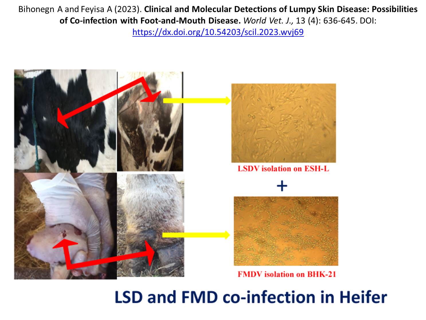
Clinical and Molecular Detections of Lumpy Skin Disease: Possibilities of Co-infection with Foot-and-Mouth Disease
Bihonegn A and Feyisa A.
World Vet. J. 13(4): 636-645, 2023; pii:S232245682300069-13
DOI: https://dx.doi.org/10.54203/scil.2023.wvj69
ABSTRACT: Lumpy skin disease (LSD) and foot and mouth disease (FMD) are notable viral diseases of cattle. This report aimed to highlight the possibilities of an uncommon case of LSD and FMD co-infection. The report also presents the clinical and molecular detection of LSD virus in six crossbred calves and LSD and FMD virus co-infection in a heifer at small-scale dairy farms located in northern Ethiopia. Nasal swabs and tissue samples were collected following aseptic techniques from the six calves suspected of having LSD and a tissue sample from one heifer suspected of having LSD-FMD co-infection and submitted to the laboratory for cell culture and real-time polymerase chain reaction (PCR) tests. Different-sized, firm, painful skin nodules with necrotic centers were seen on different parts of the calves’ body. Swelling of the prescapular and prefemoral lymph nodes, conjunctivitis, and corneal cloudiness were also observed. Uniquely, one heifer was seen with erosive lesions in the oral cavity and tongue, salivation, lameness, and skin nodules. Intracytoplasmic inclusion bodies, a distinctive feature of LSD virus, and the formation of syncytia, a characteristic of FMD virus, were observed in the cell lines. The heifer was diagnosed with a rare co-infection of LSDV and FMDV based on clinical signs, cell culture, and real-time PCR test results. The other six calves were diagnosed with the LSD virus. Treatment with broad-spectrum antibiotics, local wound cleansing, and anti-inflammatory drugs was initiated. Unfortunately, the heifer with LSD-FMD co-infection died while under treatment, and just three calves with LSD were recovered. It can be concluded that vaccination of animals against both diseases and promotion of bio-security protocols in farms is more helpful than treatment, and early case reporting is also warranted to avoid losses related to the diseases.
Keywords: Calves, Co-infection, Foot and mouth disease, Heifer, Lumpy skin disease
[Full text-PDF] [Crossref Metadata] [Scopus] [Export from ePrints]
Previous issue | Next issue | Archive
![]() This work is licensed under a Creative Commons Attribution 4.0 International License (CC BY 4.0).
This work is licensed under a Creative Commons Attribution 4.0 International License (CC BY 4.0).


